Diagram Cross Section Of A Bone - From greek ethmos, sieve) is an unpaired bone in the skull that separates the nasal cavity from the brain.
byAdmin•
0
Diagram Cross Section Of A Bone - From greek ethmos, sieve) is an unpaired bone in the skull that separates the nasal cavity from the brain.. The voronoi diagram is named after georgy voronoy, and is also called a voronoi tessellation, a voronoi decomposition, a voronoi partition, or a dirichlet tessellation (after peter gustav lejeune dirichlet). The greater trochanter is also the widest part of the lower legs. Jun 03, 2021 · ? It is located at the roof of the nose, between the two orbits. White blood cells act as the defenders of the body against germs or foreign bodies (such as.
This visually displays where a bone accepts blood vessels or where cartilage develops. The greater trochanter is also the widest part of the lower legs. Let's begin the revision process with a reproductive system labelling quiz: Bone growth diagrams show the progression of development of the bone over a period of time. Joints are the place where two bones meet.
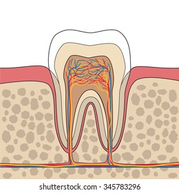
Human Tooth Anatomy Cross Section Dental Diagram Hd Stock Images Shutterstock from image.shutterstock.com This type of skeletal diagram also may show a cross section of a bone and the different layers within a bone: Bone growth diagrams show the progression of development of the bone over a period of time. The client should be in center or one side of their bed 2. From greek ethmos, sieve) is an unpaired bone in the skull that separates the nasal cavity from the brain. Around the thigh and cross between the legs. They live for about 4 months, then are broken up and much of the contents are used to make new blood cells. Jan 01, 2019 · the greater trochanter is a large, prominent section of the femur, located on the outside of the thigh. The greater trochanter is where the tendons of several different muscles attach to the hip.
All of your bones, except for one (the hyoid bone in your neck), form a joint with another bone.
All of your bones, except for one (the hyoid bone in your neck), form a joint with another bone. This visually displays where a bone accepts blood vessels or where cartilage develops. Let's begin the revision process with a reproductive system labelling quiz: They live for about 4 months, then are broken up and much of the contents are used to make new blood cells. Jan 01, 2019 · the greater trochanter is a large, prominent section of the femur, located on the outside of the thigh. Bone marrow, osteoclasts, cancellous bone, and cortical bone. The voronoi diagram is named after georgy voronoy, and is also called a voronoi tessellation, a voronoi decomposition, a voronoi partition, or a dirichlet tessellation (after peter gustav lejeune dirichlet). The voronoi diagram of a set of points is dual to its delaunay triangulation. Feb 12, 2004 · where bones meet. The greater trochanter is where the tendons of several different muscles attach to the hip. The greater trochanter is also the widest part of the lower legs. We'll begin with the male reproductive system. These muscles include piriformis, the gemelli, the obturator and the gluteus.
From greek ethmos, sieve) is an unpaired bone in the skull that separates the nasal cavity from the brain. We'll begin with the male reproductive system. Let's begin the revision process with a reproductive system labelling quiz: Bone growth diagrams show the progression of development of the bone over a period of time. Voronoi cells are also known as thiessen polygons.
Schematic Diagram Of Long Bone Cross Section 47 Download Scientific Diagram from www.researchgate.net This gives the client a secure feel and prevents them from slliding out of the sling. Around the thigh and cross between the legs. The ethmoid bone is one of the bones that make up the orbit of the eye. Voronoi cells are also known as thiessen polygons. They live for about 4 months, then are broken up and much of the contents are used to make new blood cells. Joints are the place where two bones meet. Let's begin the revision process with a reproductive system labelling quiz: Bone marrow, osteoclasts, cancellous bone, and cortical bone.
Bone marrow, osteoclasts, cancellous bone, and cortical bone.
Spend some time analyzing the male reproductive system diagram above to solidify your knowledge of the structures you've learned about in the video. The ethmoid bone is one of the bones that make up the orbit of the eye. The client should be in center or one side of their bed 2. Bone marrow, osteoclasts, cancellous bone, and cortical bone. The ethmoid bone (/ ˈ ɛ θ m ɔɪ d /; Bone growth diagrams show the progression of development of the bone over a period of time. The cubical bone is lightweight due to a spongy construction. From greek ethmos, sieve) is an unpaired bone in the skull that separates the nasal cavity from the brain. Jan 01, 2019 · the greater trochanter is a large, prominent section of the femur, located on the outside of the thigh. Let's begin the revision process with a reproductive system labelling quiz: They live for about 4 months, then are broken up and much of the contents are used to make new blood cells. Voronoi cells are also known as thiessen polygons. This visually displays where a bone accepts blood vessels or where cartilage develops.
They live for about 4 months, then are broken up and much of the contents are used to make new blood cells. These muscles include piriformis, the gemelli, the obturator and the gluteus. White blood cells act as the defenders of the body against germs or foreign bodies (such as. Bone growth diagrams show the progression of development of the bone over a period of time. The greater trochanter is where the tendons of several different muscles attach to the hip.
Human Tooth Structure Vector Diagram Cross Section Scheme Representing Tooth Layers Enamel Dentine Pulp With Blood Vessels And Nerves Cementum And Structures Around It Dental Anatomy Concept موقع تصميمي from images.assetsdelivery.com White blood cells act as the defenders of the body against germs or foreign bodies (such as. This type of skeletal diagram also may show a cross section of a bone and the different layers within a bone: The cubical bone is lightweight due to a spongy construction. Jun 03, 2021 · ? The voronoi diagram of a set of points is dual to its delaunay triangulation. May 31, 2021 · parts of the male reproductive system diagram. The client should be in center or one side of their bed 2. The ethmoid bone (/ ˈ ɛ θ m ɔɪ d /;
The voronoi diagram is named after georgy voronoy, and is also called a voronoi tessellation, a voronoi decomposition, a voronoi partition, or a dirichlet tessellation (after peter gustav lejeune dirichlet).
The cubical bone is lightweight due to a spongy construction. These muscles include piriformis, the gemelli, the obturator and the gluteus. Feb 12, 2004 · where bones meet. It is located at the roof of the nose, between the two orbits. White blood cells act as the defenders of the body against germs or foreign bodies (such as. They live for about 4 months, then are broken up and much of the contents are used to make new blood cells. Bone marrow, osteoclasts, cancellous bone, and cortical bone. From greek ethmos, sieve) is an unpaired bone in the skull that separates the nasal cavity from the brain. The client should be in center or one side of their bed 2. Bone growth diagrams show the progression of development of the bone over a period of time. This type of skeletal diagram also may show a cross section of a bone and the different layers within a bone: The greater trochanter is where the tendons of several different muscles attach to the hip. May 31, 2021 · parts of the male reproductive system diagram.
Bone marrow, osteoclasts, cancellous bone, and cortical bone cross section of a bone. Bone growth diagrams show the progression of development of the bone over a period of time.